These drops will
probably blur your vision so that you may be unable to read without
appropriate spectacles (or a magnifying lens) for up to four hours.
Remember also that you would not be able to drive a car until you are
once again able to read a number plate from a distance of 25 yards.
If your tumour is
located on the iris, the pupils will not be dilated until the consultant
has examined you.
The nurse can store
your luggage in a cupboard, if you wish. However, the hospital can not
be held responsible for any loss or theft, so please keep any valuable
items with you.
Examination by
Specialist Registrar
After seeing the sift nurse, you will be seated in the waiting area
again until you are called to see the specialist registrar, who will:
We are interested
in how your tumour was detected and how your condition was managed prior
to your referral to our Centre. This is because we are conducting a
study into the detection of ocular tumours in the community. We hope
this investigation will in future result in earlier diagnosis and treatment.
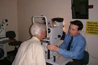
Photography
After seeing the specialist registrar, you again wait outside the
consulting room until you are called for photography. The photographer
will check your name and age before asking you to position yourself
at the camera, seated with your chin resting on the chin-rest and your
forehead pressed against a bar. Please try to keep your eye wide open
while the photographs are being taken.
Your photographs
will be used:
If any close relatives
or friends have come with you to the hospital they are welcome to accompany
you during your consultation, if you wish.
A model eye will
be used to help you understand the structure of the eye and you will
also be shown any ultrasound images of your tumour.
You are of course
encouraged to ask questions, although these are best left to the end
of the examination.
An audio-cassette
tape recording of your consultation will be given to you to help you
remember what was said. Most patients seem to find this very useful
and are quite surprised by the amount of information they missed the
first time.
Discharge from
Clinic
If on the basis of size and appearance your tumour is considered to
be benign (ie, not cancerous and therefore relatively harmless) and
if it does not require treatment, you will be discharged from the clinic.
A letter will be
written to the consultant ophthalmologist at your home hospital describing
the clinical findings, stating the diagnosis and advising on future
care.
Copies of the letter
will be sent to your general practitioner and your optician (if you
have given us your consent and informed us of the name and address).
It may be necessary
for you to return to our Centre for examination after several months,
in which case we will give you an appointment sheet. This should be
taken to the reception desk, where a specific date for your next appointment
will be selected, using the hospital computer system. Finally, you will
be given an appointment card by the receptionist.
Treatment Selection
If you
require treatment, then all the therapeutic options will be discussed,
together will treatment schedules, possible side effects, and likely
outcomes.
You will be helped
to select the best treatment for your particular condition. If possible,
a decision is made regarding treatment by the end of your consultation,
but you would still have the opportunity of changing your mind.
If you feel that
you require more time to reach a decision, this is quite possible, of
course.
Counselling
by Nurse
After your consultation you will be taken to a quiet room, where a nurse
will go over what was said, answering any more questions that might
come to mind.
If you would like
to speak to another patient who has previously received the same treatment
as yourself then the nurse would be able to arrange for you to speak
to this volunteer by telephone.
Screening for
metastatic melanoma
Before treatment of an ocular melanoma, screening consists of: