|
|
|
The word 'tumour' merely means a lump, which may be a growth or another
kind of mass, such as a blood clot or cyst.
|
| |
|
A neoplastic tumour
is a growth consisting of abnormal, new tissue. This can be benign or
malignant. A benign tumour damages the eye without posing any threat
to life. A malignant tumour can be lethal because of spread to vital
tissues around the eye and dissemination to other parts of the body
(i.e., metastasis).
Adenoma and adenocarcinoma
These are very rare tumours arising from particular membranes inside
the eye (i.e., retinal pigment epithelium and ciliary epithelium). They
can be benign or malignant. If malignant, they can invade local tissues
within and around the eye but do not usually spread to other parts of
the body.
|
 CHRPE
CHRPE
|
|
Congenital hypertrophy
of the retinal pigment epithelium
This lesion, which is also called 'chirpy', is a flat birthmark at the
back of the eye. It is a large, dark, black spot, typically with a few
white spots and discrete edges, often surrounded by a white 'halo'.
Despite its striking appearance, it is entirely harmless and does not
require any treatment. CHRPE Cyst A hollow swelling, filled with fluid.
This tends to arise behind the iris, pushing the iris forwards. Intraocular
cysts are not usually neoplastic and do not threaten life.
|
| |
|
Disciform lesion
This is a collection of fresh blood, clotted blood and scar tissue beneath
the retina. It usually arises in more elderly individuals and is caused
by abnormal veins growing beneath the retina.
|
|
|
|
Haemangioma
This is a benign tumour consisting of abnormal blood vessels. Fluid
leaking from the tumour collects beneath the retina, causing distorted
vision and blurred vision. If the amount of fluid beneath the retina
is excessive and if the retina becomes totally detached, then abnormal
blood vessels can develop on the iris. These can block the outflow of
fluid from the eye to cause an increase in pressure, which can be painful.
|
 Choroidal
haemangioma
Choroidal
haemangioma |
|
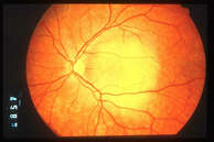
Choroidal haemangioma
The circumscribed variety usually becomes noticeable in middle age.
A diffuse variety occurs in younger patients as part of the Sturge Weber
Syndrome, which is characterized by a red birthmark on the skin of the
face.
|
| |
|
Leiomyoma
A very rare benign tumour consisting of muscle.
Melanoma
This is a malignant tumour arising from melanocytes. Intraocular melanoma
develops within the choroid, ciliary body or iris. Extraocular melanoma
develops in conjunctiva or skin.
|
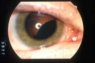
 Iris
melanoma
Iris
melanoma
|
|
Iris melanoma
Intraocular (or 'uveal') melanomas affect about one person in every
2500 whereas conjunctival melanomas affect one person in every 125,000.
Both tumours tend to affect adults. The cause is unknown, although as
with skin melanomas, ocular melanomas tend to be more common in individuals
with fair skin, light-coloured eyes, and a tendency to sunburn.
|
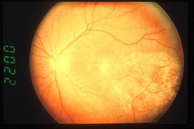
 Colour
photograph of the back
Colour
photograph of the back
of the left eye showing
(a) choroidal melanoma,
(b) optic nerve, and (c) fovea.
The retina is transparent except for
its branching arteries and veins
•
|
|
Choroidal melanoma
tends to:
- Cause retinal
detachment, with blurred vision, distorted vision, flashes of light
and a visual field defect;
- Perforate the retina,
to cause vitreous haemorrhage (i.e., bleeding into the jelly), with
floaters and blurred vision;
- Grow through the
sclera (i.e., wall of the eye) to invade the tissues around the eye.
|
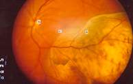
 Ciliary
body tumour
Ciliary
body tumour |
|
Ciliary body melanoma
tends to:
- Press on the lens,
to displace and distort the lens, which also becomes cataractous (i.e.,
cloudy) to cause distorted vision and blurred vision.
- Invade the iris
to become visible to the naked eye;
- Invade the gutter
draining fluid from the eye (i.e., trabecular meshwork in angle of anterior
chamber) to cause raised intraocular pressure and loss of vision (i.e.,
secondary glaucoma).
- Spread to tissues
around the eye.
|
| |
|
Ciliary body tumour
Iris melanomas tend to:
- Press on the lens
to cause cataract;
- Invade the angle
to cause glaucoma.
Conjunctival melanoma
tends to:
- Form a nodule
in the transparent conjunctiva over the white of the eye or on the inner
surface of the eyelid;
- Spread in a diffuse
fashion in the conjunctiva;
- Scatter tumour
cells to glands in the cheek and neck, where new tumours may develop;
- Form new tumours
in other parts of the body.
Sooner or later,
melanomas develop the capacity to scatter malignant cells to distant
parts of the body, particularly the liver. This process is called metastasis.
Intraocular melanomas metastasize via the blood circulation whereas
conjunctival melanomas can also spread through the lymphatic system.
|
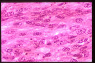 Microscopic
appearance of spindle
Microscopic
appearance of spindle
cell melanoma. Cells are long and narrow and tightly packed together.
|
|
With intraocular
melanomas, the chances of metastasis depend on:
- The size of the
tumour;
- Transformation
of tumour cell shape from spindle (i.e., spiky) to epithelioid (i.e.,
round);
- The formation
of circular membranes within the tumour (i.e., closed loops);
- The loss of inhibitory
controlling genes within the tumour, particularly those on chromosome
3;
- Tumour spread
outside the eye;
- Tumour recurrence
after radiotherapy or resection.
|
|
|
|
With extraocular
melanomas, the chances of metastasis depend on features such as:
- The thickness
of the tumour;
- Location in the
fornix (i.e., where the eye meets the eyelid).
Tumour recurrence
after radiotherapy or resection is unlikely to be the cause of metastatic
disease but probably indicates that the original tumour was relatively
aggressive.
Most patients with
ocular melanoma have a good prognosis for survival when first seen and
treated.
|
 Choroidal
metastasis
Choroidal
metastasis |
|
Metastasis
This is a malignant tumour spreading via the blood circulation
to the eye from a cancer in another part of the body, such breast or
lung.
This tumour is
usually yellow or white. It grows rapidly and leaks large amounts of
fluid to cause progressive loss of vision. It usually responds to a
small dose of external beam radiotherapy, with improvement in vision.
|
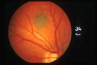 Choroidal
naevus
Choroidal
naevus |
|
Naevus (plural:
naevi)
This is a benign 'mole' arising from melanocytes. It forms a grey, brown
or yellow lump, either in the choroid, beneath the retina, or on the
iris. Choroidal naevi are very common, being present in about one in
ten individuals. They differ from malignant melanomas in that they usually:
- have a thickness
less than 2 mm;
- do not cause symptoms;
- do not leak significant
amounts of fluid;
- do not have large
amounts of 'orange pigment' on their surface.
|
|
|
|
Iris naevi
tend to be smaller than malignant melanomas, and usually not more than
3 mm in diameter.
In some patients,
the only way to be sure that a tumour is a benign naevus and not a malignant
melanoma is to observe the lesion for many years to ensure that the
tumour does not grow.
|
|
|
|
Neurilemmoma
A very rare benign tumour arising from nerve tissue.
Osteoma
A very rare tumour consisting of bone within the eye, usually next to
the optic nerve.
Retinoblastoma
This is a highly malignant tumour developing in the retina of a baby
or infant. It develops when both the chromosome 13 inherited from the
father and the chromosome 13 inherited from the mother are mutated (i.e.,
two-hit hypothesis). In some babies, both mutations occur in the same
cell, so that only one retinoblastoma develops. Other babies inherit
one mutation from a parent, so that every cell in the body is abnormal
and so that they tend to develop numerous retinoblastomas in both eyes
as well as other cancers in various parts of the body.


|