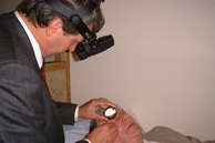
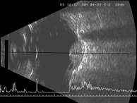
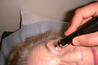
This page describes the various examinations and tests that are in common use at our clinic.


Slit-lamp examination
The slit-lamp allows a highly magnified view of the eye, with well controlled illumination allowing a clear view of the tumour. The source of light can either be diffuse or slit-like (hence the name of the instrument). It is possible to adjust the length of the slit, which can therefore be used to measure the size of a tumour.

A number of hand-held lenses are available to facilitate examination. For example, the Goldmann three-mirror lens enables the examiner to see round corners whereas the Volk 'Superfield' lens allows a wide-angle stereoscopic view of a large area of the back of the eye.

Ophthalmoscopy
There are two types of ophthalmoscopy, direct and indirect. With direct ophthalmoscopy the examiner sees the retina directly. With indirect ophthalmoscopy, a hand-held lens is used to project an image in space, a few inches in front of the eye, and it is this image that is seen by the examiner. Compared to direct ophthalmoscopy, indirect ophthal-moscopy produces a much wider field of view, which is also three-dimensional and therefore much easier to interpret.


Binocular indirect ophthalmoscopy
 
Binocular indirect ophthalmoscopy
Most tumours can be diagnosed by their appearance on ophthalmoscopy or slit-lamp examination. It may be necessary to monitor a lesion over several months or years to detect change.
Difficulties can arise if the tumour is not visible because of vitreous haemorrhage or cataract. These can be overcome by treating the cataract, waiting for the vitreous haemorrhage to clear spontaneously, or perhaps removing the haemorrhage by vitrectomy.
   

Colour photography
Colour photography is useful for documenting the appearances of the tumour so that any change over time can be readily detected. Such photography is possible with tumours extending far back in the eye, near the fovea and optic disc. Tumours closer to the front of the eye, however, are inaccessible to most retinal cameras. Newer equipment is being developed, which promises to overcome this problem.


Fluorescein angiogram
 
Angiography
Angiography is performed by injecting a dye into a vein in the arm and then taking a succession of photographs of the back of the eye.
   

The dye is fluorescent to that it has the property of changing light from one colour to another. The photographs are taken using a flash and a filter of the appropriate colours. The dye can therefore be seen shining brightly as it passes through the arteries and veins and as it leaks through any abnormal areas. There are two kinds of angiography: fluorescein angiography and indocyanine green angiography.

With fluorescein angiography, blue light is transformed by the dye into green light.

The blue light does not penetrate very deeply into the ocular tissues and therefore provides information only on the surface structures, such as the retina, the retinal pigment epithelium and perhaps any exposed tumour.

Fluorescein angiogram
The injected fluorescein dye tends to cause yellowing of the skin and urine for a few hours and about one in ten patients experience transient nausea, although vomiting is rare. About one in 2000 patients develops a severe anaphylactic (ie, allergic) reaction, with collapse. About one in two hundred thousand patients is believed to die as a result of fluorescein injection.

The principles of indocyanine green angiography are similar to fluorescein angiography except that red light is used, which penetrates more deeply into tissues.


Mr Damato performing
ultrasonography with the assistance
of Sister Kathy Mulhearn


Ultrasound probe in position


Ultrasonography
With ultrasonography, high-frequency, inaudible sound waves are emitted into the eye. These waves bounce off any tissue surface back towards the probe, which measures the 'loudness' of the reflected sound and the time taken for the sound to travel into the eye and back again. The intensity of the reflected signal gives an idea of the 'hardness' of the reflecting tissue. The time taken for the reflected signal to be received gives an indication of the distance travelled by the sound.

A-scan ultrasonography produces a linear signal, with a series of waves, which reveal the consistency of the tumour.


B-Scan ultrasound
 
With B-scan ultrasonography, the beam sweeps the eye from side to side, producing a visual slice of the eye and a good idea of the size and shape of any tumour in the eye.
   

Ultrasonography has several applications in assessing an eye with a tumour:

  • If the media are opaque, it enables the tumour to be detected.
  • Ultrasonography can also reveal tumour extension outside the eye.
  • By demonstrating a collar-stud shape, ultrasonography can help establish the diagnosis.
  • With calipers, it is possible to measure tumour thickness and basal diameter. These measurements are useful when selecting treatment and measuring how a tumour is growing or regressing over time. Care must be taken, however, to ensure that any repeated measurements are performed in a consistent manner.

Tumours close to the front of the eye are difficult to measure with conventional ultrasonography. Special high-frequency probes have recently been developed, which provide very clear views of the anterior chamber of the eye. These require the use of a small eye-bath, filled with hypromellose, which is a viscous, clear fluid.

Magnetic Resonance Imaging
Magnetic resonance imaging is performed by emitting pulses of magnetism through the body so that all the atoms spin in the same direction thereby giving rise to electrical fields, which can then be measured and converted into images. This type of scan produces very clear pictures of the eye, with different tissues showing different degrees of brightness. Melanin, for example, becomes bright with one type of MRI scan (ie, 'T1') and dark with another type (ie, 'T2'). Although this information is helpful in certain circumstances, there are limitations, because not all melanomas have melanin and, conversely, not all melanotic tumours are melanomas.

Magnetic resonance imaging is expensive and there may be a waiting list. Furthermore, the examination can be quite stressful if the patient suffers from claustrophobia. For these reasons, it is not performed routinely but is only reserved for the rare instances when the diagnosis is not provided by ophthalmoscopy and ultrasonography.

Computerized tomography
CT scans are obtained by passing very fine x-rays through the body from different directions and then reconstructing the results to create an image 'slice' of the body. This type of scan does not usually provide more information than ultrasonography, which is more convenient and less expensive.

Biopsy
The large majority of intraocular tumours can be diagnosed quite reliably by ophthalmoscopy and ultrasonography. Biopsy is useful for the rare instance when there is considerable doubt about the diagnosis despite full clinical examination.

Fine needle aspiration biopsy
Fine needle aspiration biopsy (FNAB) is performed by passing a very fine needle through the eye into the middle of the tumour and then gently moving the tip of the needle backwards and forwards a few times so that tiny tumour fragments are forced up the needle. Gentle suction can be applied at the same time to improve the yield.

Fine needle aspiration biopsy has the advantage of being a simple procedure from the surgical point of view. However, the yield of tumour cells is small and may not be enough to allow the diagnosis to be confirmed by special stains. To ensure that an adequate specimen has been obtained it is helpful if the pathologist can come to the operating theatre suite equipped with a microscope.

One might imagine that passing a needle through the retina would inevitably cause a retinal detachment, but this complication is surprisingly rare. There is often a mild haemorrhage, which can cause blurred vision or floaters, but this usually resolves spontaneously in a short time.

Incisional biopsy
Incisional biopsy is performed by creating a small trapdoor directly over the tumour and removing a small sample with scissors. This is a more difficult procedure than fine needle aspiration biopsy, and is usually performed under general anaesthesia, with mild or moderate lowering of the blood pressure.

There is also a risk of seeding tumour cells into the normal tissues around the eye and if the tumour is not treated quickly it may spread through the opening created by the surgery. For these reasons, a ruthenium plaque is placed over the area of the biopsy during the same procedure, selecting the time for which the plaque is left in place according to the diagnosis of the tumour. For example, if pathological examination shows the tumour to be a metastasis then the plaque is removed after delivering a dose of about 40 Gy to the tumour summit whereas if it is a melanoma, the exposure time is twice as long.

Excisional biopsy
Excisional biopsy involves total removal of the tumour, thereby providing both a diagnosis and a cure. It is mostly performed if local resection would be the treatment of choice in any case. Occasionally, if the eye is blind, painful and painful or unsightly the most practical solution is to remove the eye and to establish the diagnosis by pathological examination.