This page gives a brief overview of the various methods available for treating ocular tumours. More detailed information will be given to you once the most appropriate form of treatment for your particular tumour has been selected.


Good result after plaque radiotherapy, with black area of dead tumour
(ie, 'pile of soot') surrounded by white area.
 

Plaque radiotherapy
This treatment is indicated for selected choroidal melanomas, conjunctival melanomas, choroidal haemangiomas and metastases. It enables a high dose of radiation to be focused onto a small area and has the advantage of being completed in a few days.

The radiotherapy is administred by means of a saucer-shaped plaque, which has an inner, concave radioactive surface and an outer, convex protective shield.


The plaque is placed against the wall of the eye directly over the tumour and held in place with sutures. It is removed between two and seven days later, once the appropriate dose of radiation has been delivered. We can select between ruthenium plaques, which are suitable for tumours up to approximately 5 mm thick, and iodine plaques, which can treat tumours up to 9 mm thick (albeit giving a higher dose of radiation to normal ocular structures). Ruthenium plaques are available at short notice, whereas iodine plaques need to be constructed for each patient and this takes three or four weeks.


Plan for proton beam radiotherapy
 

Proton beam radiotherapy
This treatment is selected when the tumour is not suitable for the more convenient plaque radiotherapy, that is, small intraocular melanomas near the optic nerve, choroidal haemangiomas, iris melanomas, and some large intraocular melanomas.

The treatment involves:

  • A 45 minute operation under general anaesthesia, to suture four tiny tantalum markers to the wall of the eye, at known locations in relation to the tumour margins;
  • Treatment planning for half a day at Clatterbridge Centre for Oncology (CCO);
   
  • A five day course of radiotherapy at CCO, with each session taking about one hour.
  • An examination at your local eye hospital about eight weeks after the radiotherapy.

Stereotactic radiotherapy
This treatment involves directing radiation at the tumour from several directions so as to maximize the dose of radiation within the tumour while minimizing the radiation delivered to surrounding healthy tissues. This treatment is still experimental and not performed at our hospital.


Right eye showing choroidal melanoma before local resection (a)


Post-operative appearance showing (a) inner surface of white sclera, (b) retinal vein, and (c) small residual haemorrhage.


Trans-scleral local resection
Trans-scleral local resection involves the removal of the tumour through a large trapdoor in the wall of the eye. To prevent bleeding, the intraocular pressure is lowered, using hypotensive anaesthesia, monitoring the brain and heart using special equipment. At the end of the procedure, a radioactive plaque is placed over the treated area to reduce the chances of tumour recurrence. Each operation takes about three to four hours. This is difficult surgery and therefore performed only when an intraocular melanoma is too large for treatment with radiotherapy.


Trans-retinal local resection
 

Trans-retinal local resection
In this operation, the tumour is 'hoovered' through a hole in the retina, using a vacuum cleaner, which is about the same size as a match-stick. Laser treatment is given to prevent tumour recurrence. The eye is filled with silicone oil for about twelve weeks to hold the retina in place until scarring has firmly welded the retina in position.


This operation is difficult, semi-experimental and controversial and therefore used as a last resort when a tumour is located next to the optic nerve and when it is important to conserve vision.

Conjunctival excision
Discrete tumour nodules on the surface of the eyeball can be removed surgically, if not too extensive. This operation is usually performed under general anaesthesia.

Phototherapy
Laser treatment involves heating the tumour for about one minute, using an infrared laser beam. The treatment lasts about half an hour and is delivered under local anaesthesia on an outpatient basis.

This treatment is suitable for small tumours, which are not more than 3 mm thick, and which are too close to the optic nerve for radiotherapy.

Cryotherapy
Very thin tumours on the surface of the eyeball can be given 'freezing treatment', using either a spray of liquid nitrogen or a special pencil-like device. This treatment can be administered under local or general anaesthesia.

External beam radiotherapy
External beam radiotherapy is indicated for choroidal haemangiomas and metastases. These tumours respond to doses of radiation that are low enough to be well tolerated by the eye. The equipment required for this treatment is available at most hospitals. To reduce complications, the treatment is given in small doses over three to four weeks. The eye is treated from the side to avoid cataract.

Topical chemotherapy
Very thin tumours on the surface of the eye that are too extensive for surgical removal can be treated with special drops ('weedkiller'), consisting of Mitomycin C or 5-FU. These drops are administered on an outpatient basis, with each course lasting between two and four weeks.
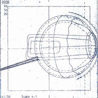
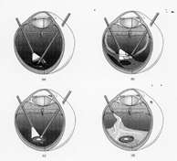
Enucleation
Removal of the eye is indicated when the chances of conserving a useful eye are low and when the risk of complications is high.

The operation is performed under general anaesthesia. A long-acting anaesthetic injection is given during the anaesthesia to minimize pain during the first few post-operative days. The enucleated eye is replaced by a bone-like ball implant. The eye muscles are sutured to this implant so that the artificial eye will move with the fellow eye. At the end of the operation a transparent 'conformer', similar to a rigid contact lens, is placed in the socket. About ten weeks after surgery, this is replaced by a 'tailor-made' permanent artificial eye, at the patient's referring hospital.

Exenteration
In very rare instances it is necessary to remove not only the eye but also the surrounding tissues and the eyelids. A special cosmetic prosthesis is made after the operation.